You are here
Leaf Structure and Function
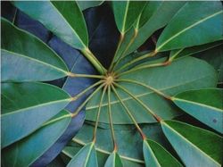
For a typical leaf, we use that of the umbrella tree, which is commonly sold as a foliage plant throughout North America and Europe. It is actually a tree native to tropical rainforests of northern Australia; it is a good example because we can examine it at any time of the year.
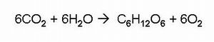
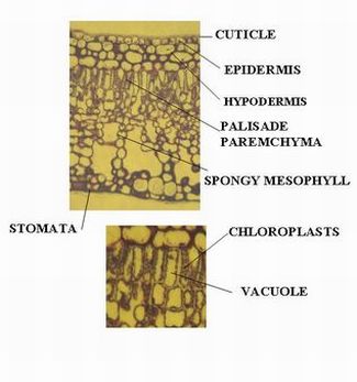
The structure of the umbrella tree leaf is typical of leaves in general (Above left photo). It has an outer layer, the epidermis, which produces a waxy waterproof coating. The epidermis of the undersurface produces guard cells, which swell and shrink to close and open the pores (stomata) which control the loss of water vapor (transpiration) and the entry of carbon dioxide. The internal tissues consist of the mesophyll, the photosynthetic cells of the leaf. These are typically the long columnar cells nearer the surface (palisade parenchyma) and the looser irregular cells beneath (the spongy mesophyll parenchyma). These cells are loaded with chloroplasts in the cytoplasm. Each of these cells has a large vacuole, bound by a membrane, which takes up some 90 % of the cell volume. Water and nutrients move into these tissues via the xylem tissue in the veins, and the sugar products of photosynthesis are translocated to other parts of the plant via the phloem tissue. The importance of the movement of carbon dioxide and water into the plant are seen in the summary equation for photosynthesis. (Cross Section in Above Right Photo)
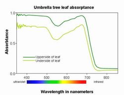
Color is produced by the balance of pigments in the leaf tissue and also by the distribution of pigments in the plastids as well as the air spaces inside of the leaf that scatter the light penetrating into the leaf. Thus the portions of the spectrum absorbed by the leaf, only bear a general relationship to the absorptance of the most abundant pigments, chlorophyll.
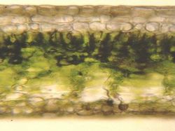
Umbrella Leaf Anatomy
The anatomy of an umbrella tree leaf, of the entire transverse section, with major tissues identified, and a detail of palisade parenchyma cells
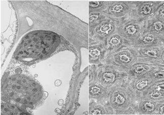
Umbrella Tree Palisade Cell and Stomata
Image on Left - Below: Transmission electron microscope photograph of the palisade parenchyma cell, showing chloroplasts with dark grana stacks and the large vacuole .
Image on Right: Scanning electron microscope photograph of the undersurface of the leaf, revealing the high density of openings (the stomata), each surrounded by two guard cells.
Umbrella Tree Absorbance